

|


| Fracture of The Heel Bone (Calcaneus) |
The ankle sprain is the most
common athletic injury. Nearly 85% of ankle sprains occur laterally,
or on the outside of ankle joints. Sprains on the inside ligaments
are less common. An ankle sprain occurs when the ankle rolls causing
the ligaments to be stretched or torn. Many sprains occur when participating
in sports, or by twisting the ankle when walking on an uneven surface.
Some individuals, due to their bone structure or foot type, are
more prone to ankle sprains. Although ankle sprains often heal without
need for surgery, it is important toconsider the problem as a partial
dislocation of the ankle joint and treat it accordingly. Ankle sprains are the most common sports injury and also the most common sports injury that presents to the emergency room. However, due to the high number of sprained ankle cases each year, they are greatly under treated. This often leads to chronic ankle instability and a feeling of looseness of the ankle joint. The loose ankle is more likely to result in constant sprains and often lead to cartilage damage and tendon tears of the ankle region. sprained ankleprotocol has been set up to assist with ankle sprains and prevent future problems. Ankle sprains are categorized according to the level of damage to the region. Prior to treatment, the type of sprain must be understood. |
| Grading ankle sprain |
Fractures of the calcaneus can be debilitating
injuries. Usually these fractures occur when tremendous forces impact
the foot and damage the heel. Examples are falls from heights or
motor vehicle accidents. Imagine standing on an orange and seeing
it widen and squash flat. This is essentially what happens to the
calcaneus. |
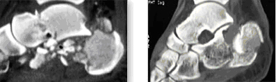 |
These are CAT scan images of fractures of the
calcaneus. The terrible injury on the left has multiple small fragments
of bone. The one on the right is more deformed but there are fewer
and larger pieces of bone. The joint between the calcaneus and the talus is called the subtalar joint. This joint is responsible for the inward and outward movements of the foot, otherwise called inversion and eversion. When the calcaneus is fractured the movement of inversion and eversion is commonly decreased or lost completely. The upward and downward movement of the ankle (dorsiflexion and plantarflexion) is not usually affected by fractures of the calcaneus. There are numerous problems associated with fractures of the calcaneus. One is the widening and deformity of the bone itself. Another is irregularity of the subtalar joint that leads to arthritis. Fractures to the calcaneus may also cause injuries to the heel cushion (the heel pad) and to the nerves and tendons surrounding the heel. |
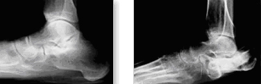 |
On the left is a picture of a foot that suffered
a bad calcaneus fracture and was treated without surgery. Compare
the shape of the flattened out heel on the left with the normal
heel on the right The ideal goal of treatment is to restore the dimensions of the heel as accurately as possible. This is always difficult because of the multiple fragments of bone that are commonly present. It is almost like trying to piece together a jigsaw puzzle. |
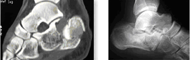 |
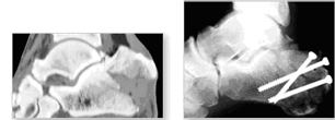
These are images of a fracture of the calcaneus.
The image on the left is a CAT scan. The image on the right is an
x-ray. This is a relatively minor fracture with only two or three
major fragments. For the majority of patients, surgery is the correct form of treatment. The goal of surgery is to restore the correct size and structure of the heel. This is done by performing what is called an open reduction and internal fixation of the fracture. The open reduction and internal fixation procedure is performed through an incision on the outside of the heel. The bone is put together and held in place with a metal plate and multiple screws. This procedure decreases the likelihood of arthritis developing and maximizes the potential for inward and outward movement of the foot. There are times, however, when the bone is so severely smashed and fractured that, in addition to the open reduction and internal fixation, the heel joint (the subtalar joint) needs to be fused. This is done to decrease the chances of developing painful arthritis. Although the inversion and eversion movement of the foot is lost after a subtalar fusion, there is a more rapid return to activities and functions after this type of surgery. |
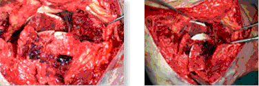 |
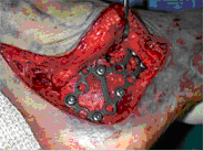
This is the inside of the foot during surgery.
There are many pieces of the calcaneus. In the right hand picture,
some of the bone pieces are shown with the white cartilage surface
broken. |
|
The fracture has been repaired with a plate
and multiple screws. Note that the overall shape of the heel (calcaneus)
has been very nicely restored. The ideal time to perform surgery is when there is minimal swelling of the skin. We will often use a foot pump device applied to the foot for a few days to decrease the swelling. This allows us to perform the surgery as soon as possible. |
 |
This fracture of the calcaneus above has been
fixed with screws that have been inserted through skin punctures
instead of large skin incisions. |
 |
The x-ray on the left shows the foot prior
to surgery. The x-ray on the right shows the foot after the procedure.
Note that the height and shape of the calcaneus have been perfectly
restored. Following surgery, no walking on the foot is permitted for approximately 3 months. A bandage is applied to the leg after surgery. After the stitches are removed, movement exercises and therapy are started to try to maximize the function of the foot. It typically takes approximately six months to recover from this type of injury. |
| ©
Foot & Ankle Associates. All Rights Reserved. |
Website Powered
by : Sterling Softwares |